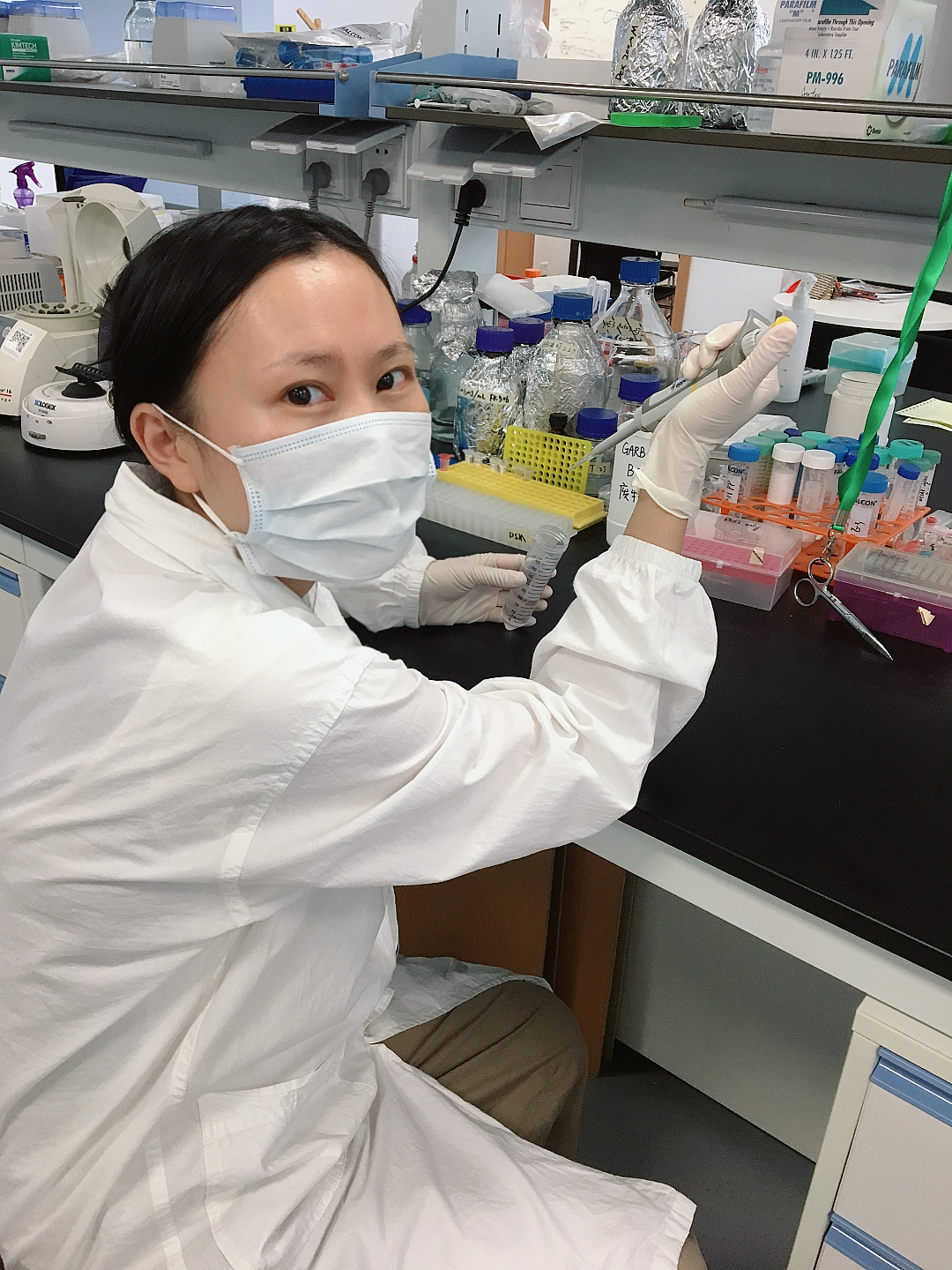
Principal investigatorName: Christopher L. AntosAssociate Professor , PhD, Associate Professor
Position: Affiliation: School of Life Science and Technology
Honor: Education Background:
Working Experience:
Group Introduction Research Area:
Heart and Appendage Regeneration
Research Interests:
The Antos group researches tissue regeneration. To understand the biology of regeneration, Dr. Antos is interested in answering three fundamental questions: Research AchievementWe are researching what electrophysiological changes occur during embryogenesis and organ regeneration and how these changes control the organ development and regeneration. Thus far, we have been determining how electrophysiological signals control how much of an organ should develop during embryogenesis and how much should regenerate after amputation injury (Dev. Cell 2014; Elife 2021; PLoS Biology 2024). In addition, we are currently examining how second messenger signals are involved in the development and regeneration of the heart (manuscript in progress). Paper Details: Jiang XW, Zhao K, Yi S, Chao Y, Xiong TL, Wang S, Yi Y, Dong SM, Chen XD, Liu R, Yan X, and Antos CL. (2024) The scale of zebrafish pectoral fin buds is determined by intercellular K+ levels and consequent Ca2+-mediated signaling via Retinoic Acid regulation of Rcan2 and Kcnk5b PLoS Biology 22(3):e3002565 Significance: The scientific significance of this paper is that it shows how retinoic acid scales vertebrate appendages through a bioelectric mechanism that controls the entire fin/embryonic fin bud/embryonic limb bud developmental programs. The technological significance of this paper is that we established a measurement/visualization method using Fluorescence Lifetime Microscopy (FLIM) with Förster Resonance Energy Transfer (FRET)-based sensor for assessing in vivo electrophysiological phenomena in three-dimensional space in developing embryo, which is not possible with current patch-clamp technologies. The specific discoveries: 1) We show that endogenous intracellular K+ levels decrease in embryonic pectoral fin buds (of live animals using 3D in vivo FLIM-FRET measurements) during developmental outgrowth, and overexpression of K+-leak channels (that decrease intracellular K+) enhance the proportions of the embryonic fin buds and adult fins by up-regulating the developmental programs of the pectoral fin buds and adult fins. Because the developmental program of the pectoral fin bud is conserved in early embryonic limb bud, our findings argue this scaling mechanism is involved in proportional growth of early fin/limb buds in all vertebrate embryos. 2) Retinoic acid decreases intracellular K+ by up-regulating rcan2 transcription. 3) Rcan2 requires the K+-leak channel Kcnk5b to enhance fin bud scaling, and it regulates Kcnk5b through serine345 in the cytoplasmic C-terminal tail of this channel. 4) Kcnk5b scales pectoral fin buds by increasing cell depolarization. 5) K+-leak channel activity requires IP3R-mediated release of Ca2+ and CaMKK to alter the scale of pectoral fin buds. Thus, we show how an electrophysiological phenomenon (K+-leak channel activity) that scales appendages is integrated into the known (retinoic acid and shh) and unknown (rcan2, kcnk5b, IP3R-mediated Ca2+ release and CaMKK regulation of shh transcription) mechanisms of early embryonic bud development. Yi C, Spitters TW, Al-Far EEA, Wang S, Xiong T, Cai S, Yan X, Guan K, Wagner M, El-Armouche A, Antos CL. (2021) A calcineurin-mediated scaling mechanism that controls a K+-leak channel to regulate morphogen and growth factor transcription. Elife 10.7554/eLife.60691 IF: 8.14 Significance: This paper shows how calcineurin scales fish fins through the post-translational modification of a serine (serine345) in the cytoplasmic tail of the K+-leak channel Kcnk5b and shows that Kcnk5b can induce different transcriptional programs depending on the cell type. Thus, this work provides an endogenous regulatory mechanism that controls cellular potassium conductance (an electrophysiological phenomenon that scales fin size) and provides an explanation for how a single type of potassium leak channel can augment the developmental growth by being able to regulate different transcriptional programs in the different tissue types of the entire anatomical structure. Lu T, Li Y, Lu W, Spitters TWGM, Wang J, Cai S, Gao J, Zhou Y, Duan Z, Xiong H, Liu L, Li Q, Jiang H, Chen K, Zhou H, Lin H, Feng H, Zhou B*, Antos CL*, Luo C* (2021) Discovery of a subtype-selective, covalent inhibitor against palmitoylation pocket of TEAD3. Acta Pharmaceutica Sinica 10.1016/j.apsb.2021.04.015 Significance: This paper addresses two points. The scientific significance is that this work indicates that different TEADs control the proportion growth of different anatomical structures of fish. We found that TEAD3 only regulates the proportional growth of the zebrafish appendages. TEAD1 appears to regulate the proportional growth of the entire body. TEADs are transcription factors that regulate Hippo signaling, a signal transduction pathway that can control the proportional growth of tissue cells. This pathway is also linked to cancer. In relation to cancer, this paper identifies small molecules that can inhibit a specific TEAD isoform (TEAD3) by interacting with features unique to this isoform, thereby limiting potential side effects of inhibiting all isoforms. Kujawski S, Lin W, Kitte F, Börmel M, Fuchs S, Arulmozhivarman G, Vogt S, Theil D, Zhang Y and Antos CL (2014) Calcineurin regulates coordinated outgrowth of zebrafish regenerating fins. Developmental Cell 28: 573-587 Significance: The importance of this work is that it provides a previously unknown mechanism for controlling the proportional growth of zebrafish appendages. Specifically. This paper shows that calcineurin acts as a molecular switch to determine whether zebrafish appendages will grow isometrically (at the same rate as the body) or allometrically (at a higher rate than the body) by controlling the entire developmental programs involved in appendage growth. It also provides evidence that calcineurin controls enhances the scale of the appendages by altering positional information of cells to make them think that they are more proximally located in the appendages than they actually are so that they grow/regenerate at a higher rate and grow/regenerate more distal structures than originally designed. Kizil C, Küchler B, Yan J-J, Özhan G, Moro E, Argenton F, Brand M, Weidinger G and Antos CL (2014) Simplet/Fam53b (Smp) is required for Wnt signal transduction by regulating β-catenin nuclear localization Development 141: 3529-3539. Significance: β-catenin-dependent Wnt signal transduction cascade is important for many physiological phenomena (stem cell biology, embryonic development, tissue regeneration, etc.). This paper identifies how the molecule Fam53b with unique conserved protein domains that have unknown function—that my research group previously showed can regulate the blastema cells (the undifferentiated progenitor cells that regenerate new tissues) in the fin, heart and possibly stem cell in the brain—is involved in maintaining β-catenin in the nucleus of cells to promote β-catenin-dependent signaling. We also identified which domains are responsible for the interaction with β-catenin and its sequestration in the nucleus. Representative Publications (*First Author, # Corresponding Author)
MonographPatentFundingAwardsResearch AchievementGroup Member and Photo
|


